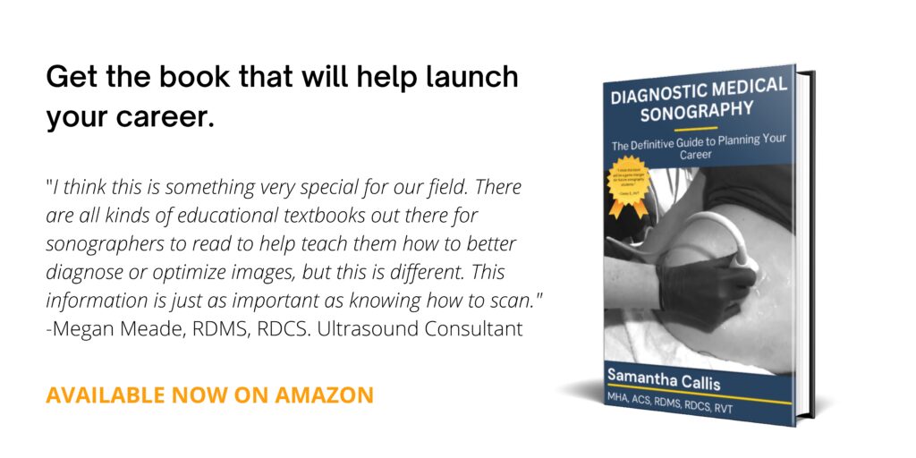
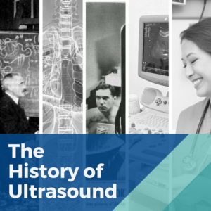
An Overview of Ultrasound History and Discovery
The technology used in medical ultrasound is continuously evolving and currently contributing to important improvments in patient diagnosis and treatment. The science and technologies employed in sonography have a long and interesting history. This story begins with the women and men (and yes animals) from across the world who have contributed to the evolution of ultrasound over the past 225+ years.
Let’s take a look back at the history of ultrasound and learn how the use of sound waves as a diagnostic tool made their way into clinics and hospitals across the globe.
Echolocation and Ultrasound’s Early Beginnings
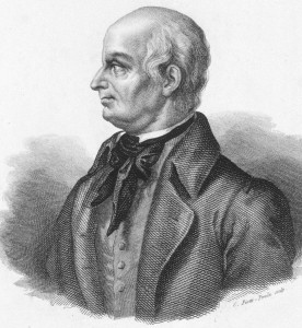
Lazzaro Spallanzani
Many ask, who invented the ultrasound? Italian biologist, Lazzaro Spallanzani is most often credited person for discovering ultrasonography.
Lazzaro Spallanzani (1729-1799) was a physiologist, professor and priest who carried out numerous experiments that led to great insights in human and animal biology.
In 1794 Spallanzani performed studies on bats that concluded that they could navigate using sound rather than sight. This is now known as echolocation where locations are determined or identified through sound waves being reflected or bounced back from objects in an environment. These same principles are how medical ultrasound technology functions today.
RELATED: 7 Female Pioneers in Medical Imaging
Ultrasound is characterized as sound waves with a frequency higher than what is audible to the human ear. “The first detailed experiments that indicated that non-audible sound might exist were performed on bats by Lazzaro Spallanzani,” states D. Kane, W. Grassi, R. Sturrock, P. V. Balint; A brief history of musculoskeletal ultrasound: ‘From bats and ships to babies and hips’, Rheumatology, Volume 43, Issue 7, 1 July 2004.
What is Echolocation?
We can find several additional examples of echolocation in nature. Echolocation pulses are short bursts of sound at frequencies that span from about 1,000 hertz in birds to at more than 200,000 hertz in whales.
Early Experiments in Ultrasound
Gerald Neuweiler, in his book The Biology of Bats, describes how Spallanzani brought owls into his lab and observed that they would not fly around the room if there was no source of light. “When he repeated the same experiment using bats, these small mammals flew confidently around the bishop’s study, even in total darkness, managing to avoid the wires that Spallanzani had hung from the ceiling,” wrote Neuweiler.
Neuweiler adds that the Italian scientist even blinded the bats by burning them with a “red-hot needle” and still they were able to avoid the wires. Spallanzani knew this because bells were attached to the ends of the wires.
The physiologist gained insight that the bats were relying on the sense of sound for navigation because when he placed closed brass tubes inside the mammals’ ears, they could not navigate the room properly and would fly into the wires.
Although he did not know that the bats were emitting their own sound for orientation, sound higher than he or any human would be able to hear, Spallanzani was able to conclude that the creatures were using their ears to navigate their environment.
Medicine Benefits from Developments in Ultrasound
As time passed, others continued to build on Spallanzani’s work. It was in 1942 that Neurologist Karl Dussik is credited with being the first to use ultrasonic waves as a diagnostic tool. He transmitted an ultrasound beam through the human skull in attempts of detecting brain tumors. This is still very early in the history of diagnostic medical sonography, but it was clear that this noninvasive technology had tremendous possibility.
Ultrasound technology and its application in healthcare have continued to mature. The advancement of tools and refinement of procedures are happening everyday. Most recently, smaller portable scanners have become more widespread, and have helped further integrate the use of ultrasound in more areas and stages of patient care.
A Chat with Sonographer, Educator, Pioneer and Ergonomics Expert, Joan P. Baker
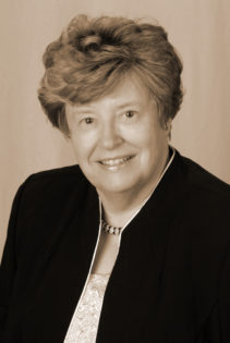
It was truly an honor to interview Joan P. Baker MSR, RDMS, RDCS, FSDMS. Originally from England, Baker was invited to the United States in the 1960s – due to her sonography passion and practice – and she’s been here ever since.
Ultrasound History Timeline
Here’s a look back at some of the key milestones in the development and history of ultrasound technology.
| Date | Historical Achievement or Event |
|---|---|
| 1794 | Physiologist Lazzaro Spallanzani was the first to study echolocation among bats, which forms the basis for ultrasound physics. |
| 1877 | Brothers Pierre and Jacques Currie discover piezoelectricity. Ultrasound transducers (probes) emit and receive sound waves by way of the piezoelectric effect. |
| 1915 | Inspired by the sinking of the Titanic, Physicist Paul Langevin was commissioned to invent a device that detected objects at the bottom of the sea. Laugevin invented a hydrophone – what the World Congress Ultrasound in Medical Education refers to as the “first transducer”. |
| 1920s-1940s | Sonography was used to treat members of European soccer teams as a form of physical therapy, to appease arthritic pain and eczema and to sterilize vaccines, states Joan Baker who holds several ARDMS ultrasound certifications. |
| 1942 | Neurologist Karl Dussik is credited with being the first to use sonography for medical diagnoses. He transmitted an ultrasound beam through the human skull in attempts of detecting brain tumors. |
| 1948 | George D. Ludwig, M.D., an Internist at the Naval Medical Research Institute, developed A-mode ultrasound equipment to detect gallstones. |
| 1949-1951 | Douglas Howry and Joseph Holmes, from the University of Colorado, were some of the leading pioneers of B-mode ultrasound equipment, including the 2D B-mode linear compound scanner. John Reid and John Wild invented a handheld B-mode device to detect breast tumors. |
| 1953 | Physician Inge Edler and Engineer C. Hellmuth Hertz performed the first successful echocardiogram by employing an echo test control device from a Siemens shipyard. |
| 1958 | Dr. Ian Donald incorporated ultrasound into the OB/GYN field of medicine. |
| 1966 | Don Baker, Dennis Watkins, and John Reid designed pulsed Doppler ultrasound technology; their developments led to imaging blood flow in various layers of the heart. |
| 1970s | The 1970s saw many developments including the continuous wave Doppler, spectral wave Doppler and color Doppler ultrasound instruments. |
| 1980s | Kazunori Baba of the University of Tokyo developed 3D ultrasound technology and captured three-dimensional images of a fetus in 1986. |
| 1989 | Professor Daniel Lichtenstein began incorporating lung and general sonography in intensive care units. |
| 1990s | Starting in the 1980s, ultrasound technology became more sophisticated with improved image quality and 3D imaging capabilities. These improvements continued into the 1990s with the adoption of 4D (real time) capabilities. Ultrasound guided biopsies (endoscopic ultrasounds) also began in the 1990s. |
| 2000s – present | Just like personal communication devices are continuously evolving and becoming more convenient, so are ultrasound technologies. A variety of compact, handheld devices have come onto the market in recent years. The iPhone now has a telesonography app and NASA has developed a virtual guidance program for non-sonographers to perform ultrasounds in space. |
History of Sonography in Obstetrics and Gynaecology
In our current culture, ultrasounds might best known for their use during pregnancy to produce a sonogram, a visual image produced from an ultrasound examination. Within the larger ultrasound family of specializations, Obstetrics and Gynaecology have seen some important historical moments as well. You’ll find some of the more notable developments in the OB/GYN specialization below.
| Date | Historical Event |
|---|---|
| 1958 | This year marked the publication of the first paper in Obstetric Ultrasound “Investigation Of Abdominal Masses By Pulsed Ultrasound” by Ian Donald, M.B.E., B.A. Cape Town, M.D. Lond., F.R.F.P.S., F.R.C.O.G. J Macvicar, M.B. Glasg., M.R.C.O.G. T.G Brown. This study marked the first ultrasound image of a fetal head. |
| 1962 – late 1960’s | George Kossoff of Australia engineers the Octason static scanner. The Octason mark 2 images allow us to see detailed fetal anatomy, and marks an important time in the development of ultrasound. |
| 1970’s | Advancements in sonography equipment and techniques progressed throughout the late 1960’s and into the 1970’s. Methods to determine the fetal biometry and fetal abnormalities continued to advance and be refined with the adaption and replacement of various techniques. |
| 1983 | Sam Maslak develops a machine that sets new standards in both spatial and contrast resolution. |
Additional information about this topic and resources consulted about the history of sonography.
If you would like to become a part of this evolving field, you can complete a degree at one of the numerous ultrasound schools across the country.